Vaccines and Anaphylaxis: Everything You Ever Wanted To Know
The Short Version: Anaphylaxis is a rapid-onset, rare manifestation of allergy which is life-threatening and involves two or more organ systems, usually including the skin and respiratory system (but not necessarily). The exact prevalence of anaphylaxis is not known but does appear to be rising (because the prevalence of allergy has been rising, for which many hypotheses have been proposed). Anaphylaxis has been documented in response to basically anything you can think of- exercise, cold air or water, heat, sunlight/ UV radiation, venom (it’s actually thought that this is how anaphylaxis evolved- animals which have anaphylactic reactions to venoms survive envenomation better), radiocontrast media (which is not the same as iodine allergy- no one is allergic to iodine), ethanol (the alcohol we drink), medications, and of course food. Even though all anaphylaxis is a medical emergency, the case-fatality ratio from anaphylaxis (despite frequent incorrect treatment) has been measured to be less than 0.001%. Anaphylaxis is foremost treated with epinephrine (though it is not unusual for people to require more than one dose from an Epipen)- despite common misconception it is NOT treated with antihistamines (though these can be given for comfort). The incidence of anaphylaxis from vaccines (excluding COVID-19 vaccines) has been measured before to be 1.31 per million vaccine doses (and in the 33 cases from that study which looked at over 25 million vaccine doses, none were fatal)- and thus is exceedingly rare. With COVID-19 vaccines, 8 cases of severe allergic reactions have been documented with just over 1 million doses doses- which makes for an incidence of approximately one per 125,000 doses of vaccine, which is still quite rare. It is not currently known what component of the vaccines is responsible, but current suspicions point to a component of the lipid nanoparticle that the mRNA is placed inside called PEG-2000. PEGs are very common additives in cosmetics, drugs, and foods, and thus allergy to them is generally very rare. Anyone receiving a vaccine is asked to stay for observation for 15 minutes in the event of a reaction, and in those who have a history of severe allergic reactions the Advisory Committee on Immunization Practices recommends observation for 30 minutes. Despite how much attention these cases of anaphylaxis have gotten in the news, anaphylaxis is readily manageable in a medical setting and still exceedingly rare.
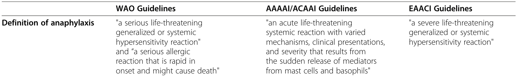
Simons FER, Ardusso LR, Bilò MB, Cardona V, Ebisawa M, El-Gamal YM, Lieberman P, Lockey RF, Muraro A, Roberts G, et al. 2014. International consensus on (ICON) anaphylaxis. World Allergy Organ J. 7(1):9. Table 2
What is Anaphylaxis?
Anaphylaxis will have a slightly different definition depending on which professional body you ask (see your right).
Adkinson NF Jr, Bochner BS, Burks DAW, Busse PWW, Holgate PST, Lemanske DRF Jr, O’Hehir PRE. 2013. Middleton’s allergy 2-volume set: Principles and practice (expert consult premium edition - enhanced online features and print). 8th ed. Philadelphia, PA: Saunders Table 77-6 summarizing the typical symptoms and signs that occur in anaphylaxis and their frequency.
Given that different professional societies use different definitions, you can surmise that anaphylaxis has a lot of complexity. The term literally means the opposite of prophylaxis, i.e. an immune response that produces illness rather than protecting you from it. Anaphylaxis can have many different symptoms, but death results from either cardiovascular collapse from the uncontrolled inflammation that results in a substantial drop in blood pressure (which is termed anaphylactic shock) or airway obstruction that prevents breathing. In addition to this, anaphylaxis usually presents with a rash (i.e. hives; but this can be delayed or not occur at all and thus its absence should not be used as an excuse to withhold therapy), as well as nausea, vomiting, diarrhea, abdominal pain, headache, dizziness, chest pain, and even heart arrhythmias in rare cases. Some patients also report a feeling of impending doom.
In general, anaphylaxis is divided into allergic and non-allergic anaphylaxis (formerly called anaphylactoid reactions; I am unsure why the term has regained use in the press recently as it is generally considered outdated). Anaphylaxis is also classified in terms of the timing patterns of the symptoms.
Castells M. 2017. Diagnosis and management of anaphylaxis in precision medicine. J Allergy Clin Immunol. 140(2):321–333. Figure 1A; Anaphylaxis can be grouped into 4 categories (endotypes). The first involves massive activation of mast cells or basophils by IgE or an IgE-independent mechanism (type 1 anaphylaxis). Sometimes certain chemotherapies and monoclonal antibodies can produce anaphylaxis reactions wherein there is massive cytokine release, and these may also present in a mixed form which resembles type 1 anaphylaxis. Complement has also been proposed as being capable of inducing anaphylaxis in response to contrast dyes, dialysis membranes, and glycosaminoglycans.
Owen J, Punt J, Stranford S. 2013. Kuby Immunology: International Edition. 7th ed. New York, NY: W.H. Freeman. Summarizing the basic mechanisms of different allergies; most allergies are due to type 1 (immediate) hypersensitivity reactions, and allergic anaphylaxis is an example.
Uniphasic- Anaphylaxis symptoms occur rapidly following exposure to the allergen, peaking within a few hours. This accounts for the vast majority of anaphylaxis cases.
Biphasic- Rarely, anaphylaxis symptoms can peak in a uniphasic pattern, but then return after an hour or two without symptoms (though up to 78 hours between symptoms has been documented), which is termed biphasic anaphylaxis. This is a rare form of anaphylaxis. Concern for this pattern of anaphylaxis means that generally anyone who experiences anaphylaxis will be observed for several hours after the episode to ensure there is no biphasic anaphylaxis.
Protracted- In some individuals the symptoms of anaphylaxis can persist without resolving for hours to days. This is uncommon and literature on this is limited.
Anaphylaxis may have no clear trigger, which is termed idiopathic anaphylaxis.
In addition, Castells also describes several endotypes (i.e. disease mechanisms) for anaphylaxis (see the figure to the right):
Type 1: IgE or non-IgE-mediated activation of mast cells and basophils
Massive cytokine release in response to certain chemotherapies and monoclonal antibodies affecting primarily T cells, monocytes and macrophages (i.e. cytokine storm-like reaction; in general however, cytokine storms are not considered a form of anaphylaxis and are not treated in the same way)
Mixed features of type 1 and cytokine release
Complement-mediated activation of mast cells or basophils
The endotype of the anaphylaxis is relevant for considerations regarding desensitization. Anaphylaxis is a manifestation of an allergic reaction, and type 1 anaphylaxis is amenable to desensitization therapy in many cases.
Allergic Anaphylaxis
Allergic anaphylaxis results from IgE antibodies that bind allergens and induce degranulation (release of contents from the granule- an organelle in the cell) from mast cells and basophils (these recognize the IgE through a receptor called FcεRI), and is thought in general to be mediated mainly by the release of large quantities of histamine; this is known as a type I (aka immediate) hypersensitivity, in more formal terms (i.e. an allergic reaction due to IgE antibodies; this classification scheme is from Gell and Coombs, see figure 15-1 at the right). IgE alone is not sufficient to cause anaphylaxis, or even any manifestation of allergy. Almost everyone (with the exception of people with certain immunological conditions) makes IgE, but relatively few people experience anaphylaxis. If you survey any given individual for their IgE, you will likely find IgE specific to a number of antigens (though in general, those with a predisposition towards allergy, called atopy, will have higher total IgE levels in their blood compared with individuals who do not have allergies) that they are not actually allergic to; the presence of IgE specific to an antigen alone (known as sensitization) DOES NOT indicate that there is an allergy to that antigen, nor does it indicate a risk of anaphylaxis (so please do not waste your money on over-the-counter food allergy tests or IgG testing; if you are concerned about a potential food allergy, please see an allergist). It is not well understood how some individuals are sensitized without apparent development of allergy. There is a vast spectrum of manifestations for type I hypersensitivities, and anaphylaxis is a rare presentation (other manifestations include hay fever, asthma, local skin eruptions, etc).
IgE must be high-affinity (i.e. it must bind tightly to the allergen) to cause degranulation that results in allergic anaphylaxis, but these IgE antibodies are not universally present. High affinity IgE production appears to be mediated by a unique, rare cell type called the Tfh13 cell (T follicular helper 13 cell, named for the IL-13 they produce), and this occurs via a different pathway than low-affinity IgE; deletion of Tfh13 cells abolished anaphylaxis in animal models. Previous studies have noted that blocking the action the cytokines produced by these cells can improve allergic disorders.
Gowthaman U, Chen JS, Zhang B, Flynn WF, Lu Y, Song W, Joseph J, Gertie JA, Xu L, Collet MA, et al. 2019. Identification of a T follicular helper cell subset that drives anaphylactic IgE. Science. 365(6456):eaaw6433 summary figure. T follicular helper cells (Tfhs) are important partner cells in the production of antibodies and guide B cells in terms of the type of antibodies they make. Tfh cells can induce different types of antibodies depending on the type of antigen they encounter. A population of Tfh cells called Tfh13 cells produces high levels of IL-4, IL-5, and IL-13, and low levels of IL-21, which seem to induce B cells to generate high-affinity IgE that drives anaphylaxis.
The exact pathophysiologic mechanisms of anaphylaxis in humans are not well understood because of difficulties in studying it. Most of the data is based on animal models, which makes extrapolation tricky, although in the past experiments were conducted in which volunteers inhaled histamine to see whether it became harder to breathe, which is kind of funny to think about (also, it absolutely does, don’t try it). Mast cells and basophils are the principal cells associated with anaphylaxis as these express FcεRI and thus are responsive to high-affinity IgE and degranulate in response to complexes of IgE and allergen. However, in addition to these, neutrophils have been recognized as playing important roles in anaphylaxis. Neutrophils can also be a source of histamine (though not as much of it), and their granules contain the myeloperoxidase (MPO) enzyme, whose levels are correlated directly with anaphylaxis severity (but the exact significance of MPO in the disease process is not well understood). Macrophages and monocytes are also thought to play a role, but it is not very well understood in human anaphylaxis, beyond production of PAF (elaborated upon shortly).
Adkinson NF Jr, Bochner BS, Burks DAW, Busse PWW, Holgate PST, Lemanske DRF Jr, O’Hehir PRE. 2013. Middleton’s allergy 2-volume set: Principles and practice (expert consult premium edition - enhanced online features and print). 8th ed. Philadelphia, PA: Saunders Table 77-4 describing the different mast cell and basophil mediators and their role in anaphylaxis.
Though histamine is generally regarded as being the major player in causing the symptoms of anaphylaxis (including wheezing, rash, diarrhea, abdominal pain, runny nose, and drops in blood pressure), it is not the only one and it is probably not even the most important one in terms of survival, as treatment with antihistamines does NOT stop anaphylaxis (but they can be given as an adjunct therapy for the patient’s comfort). Many mediators other than histamine are thought to be important in anaphylaxis however. Among these are short-lived, fat-derived molecules called autacoids, specifically platelet-activating factor (PAF, aka 1-0-alkyl-2acetyl-sn-glycero-3-phosphocholine, AGEPC, or PAF-acether) and cysteinyl-leukotrienes (CysLTs, which include LTB4, LTC4, and LTD4). PAF is made by many cells of the immune system (some lines of evidence suggest that in anaphylaxis most of it is from monocytes and macrophages) and has many diverse functions, not all of which are related to the immune system (for example, in the ovaries it helps with ovulation). PAF is so-named because it promotes aggregation, adherence, and activation of platelets, but in addition to this PAF seems to promote histamine release, and on injection produces wheal-and-flare skin lesions. Additionally, PAF levels correlate with anaphylaxis severity, and defects in PAF clearance (e.g. loss-of-function mutations in PAF acetylhydrolase, the enzyme that breaks down PAF) resulted in more severe anaphylaxis as well. Combined inhibition of PAF and histamine seems to block anaphylaxis almost completely in animals. CysLTs also produce wheal-and-flare lesions on injection, and if inhaled, produce bronchoconstriction at levels 1000 times lower than that of histamine (these are also thought to play a role in asthma).
Busse PJ, Christiansen SC. 2020. Hereditary angioedema. N Engl J Med. 382(12):1136–1148.The mechanism by which bradykinin causes angioedema. Mannose-binding lectin associated serine protease 1 (MASP1) is able to cleave the complex of high molecular weight kininogen and kallikrein (formed from prekallikrein that is cleaved by activated factor XII) to release bradykinin. Bradykinin, then, via the bradykinin B2 receptor can go on to induce vasodilation that promotes vascular leakage; the role of the B1 receptor in this process is not as well understood. Bradykinin can be inhibited with the drug icatibant, which binds B2 receptors to block bradykinin signaling. Additionally, bradykinin is broken down by ACE into des-Arg-bradykinin which is a generally inactive product but can weakly bind the B1 receptor. Bradykinin is also broken down by carboxypeptidase N and M. C1 inhibitor blocks the synthesis of bradykinin at multiple points in the synthetic pathway.
In addition to these, the complement system is thought to play a role in anaphylaxis. The complement system is a group of proteins that have been compared to tiny bombs in the blood- they can be activated by several other proteins, including IgG antibodies, and ultimately form pores in membranes which are lethal to the cells they target. In addition to these, the fragments of some complement proteins are known as anaphylatoxins; they are able to help recruit the cells of the immune system and promote leakage of the vasculature, enhancing the expression of cell adhesion molecules, and recruiting the cells of the immune system. Following anaphylaxis, complement levels are frequently depleted and in mouse models, depletion of complement before induction of anaphylaxis seems to be able to prevent it. Additionally, injection of anaphylatoxins immediately produces wheal-and-flare skin lesions at the injection site. Mast cell granules also contain a protease called tryptase which is thought to be able to induce anaphylatoxin production as well, and serum tryptase levels can aid in diagnosis of anaphylaxis as they are markers of mast cell and basophil activation (elevated tryptase levels is associated with anaphylaxis, but in pediatric patients and in food-triggered anaphylaxis, tryptase levels can be normal in anaphylaxis; the functional importance of tryptase in anaphylaxis is not entirely clear). Generally however, the role of complement in anaphylaxis is thought to be redundant with that of other proteins.
However, the complement system is also linked to the coagulation cascade and the kinin-kallikrein system, which are themselves interconnected through the intrinsic coagulation cascade. Complement activation can result in pore formation that releases bradykinin; bradykinin is a small peptide that has functions similar to histamine, and is associated with angioedema (swelling in the lower layers of the skin or mucous membranes caused by leakage of fluid from the vasculature). Bradykinin-induced angioedema often occurs in the upper airways and can be brought on by the use of ACE inhibitors (ACE is an enzyme that breaks down bradykinin) or from a deficiency of the C1 inhibitor protein of the complement system (which blocks the pathway that results in the synthesis of bradykinin as shown on the right). This is discussed further in the section on non-allergic anaphylaxis. Damage to the vasculature from the complement system can also induce the contact-dependent coagulation pathway, and induce clotting that depletes the bradykinin precursor high-molecular weight kininogen (HMWK) and factors V and VII which can produce a rare but life-threatening clotting abnormality called disseminated intravascular coagulation. Factor XII can form a complex with prekallikrein and HMWK, which forms active kallikrein that then liberates bradykinin from HMWK. Kallikrein also enhances synthesis of activated factor XII, forming a positive feedback loop. This process can be aided by the presence of negative charges which function as a scaffold and can initiate clotting in the intrinsic pathway, such as those present on heparin, which is released from mast cell and basophil granules.
Subramanian H, Gupta K, Ali H. 2016. Roles of Mas-related G protein–coupled receptor X2 on mast cell–mediated host defense, pseudoallergic drug reactions, and chronic inflammatory diseases. J Allergy Clin Immunol. 138(3):700–710 Figure 3 demonstrating the putative mechanism by which MRGPRX2 signaling occurs and can mediate pseudo-allergic reactions including non-allergic anaphylaxis. In general, binding of various substrates seems to induce import of calcium through calcium release–activated calcium channel (ORAI) which then triggers multiple downstream signaling pathways that result in mast cell degranulation.
The existence of allergic anaphylaxis that is mediated by non-IgE (i.e. IgG) antibodies is controversial in humans. Evidence for it has never been definitively demonstrated but it has been observed in other animals. There are other human allergic disorders mediated by non-IgE antibodies e.g. atopic dermatitis, but this has not been observed for anaphylaxis specifically. In general however, for these forms of anaphylaxis a much higher dose of allergen is required (which is likely because IgE binds its Fc receptors much more tightly than does IgG) and it is generally thought to occur from administering the allergen parenterally (i.e. not by mouth). In general, if a patient demonstrates IgG against an allergen, this indicates a tolerance for it.
It is also technically allergic anaphylaxis if FcεRI signaling is induced, even if IgE is not involved. Some medications are able to do this, for instance, NSAIDs.
Non-Allergic Anaphylaxis
Gao Y, Han Y, Zhang X, Fei Q, Qi R, Hou R, Cai R, Peng C, Qi Y. 2020. Penicillin causes non-allergic anaphylaxis by activating the contact system. Sci Rep. 10(1):14160. Figure 7
Non-allergic anaphylaxis refers to anaphylaxis brought on by a non-immunologic mechanism. In this case, it is thought that there are substances that are able to directly activate mast cells and induce release of granule contents that reproduce anaphylaxis or other allergic symptoms, i.e. without involving FcεRI (this is also sometimes called a pseudoallergy). This has been reported in response to exercise (which typically has a food-related trigger but not always), UV radiation/sunlight, cold air, water, heat, and many medications among many other diverse triggers. For example, non-allergic anaphylaxis to the anticoagulant heparin has been documented before, which is thought to be mediated by high levels of bradykinin, rather than histamine. NSAID medications can provoke non-allergic anaphylaxis because they block production of prostaglandin E2, which suppresses anaphylactic mediators. Vancomycin, an antibiotic, has been reported to be able to directly activate mast cells to induce histamine release, and opioid medications also appear able to provoke anaphylaxis through an unclear mechanism that directly involves opioid receptors (in the case of opioids, administration of an opioid receptor antagonist e.g. naloxone also resolves anaphylaxis). Several drugs including nicotinic receptor antagonist nonsteroidal neuromuscular blocking agents and fluoroquinolone antibiotics have been shown to be able to directly activate mast cells through a protein called Mas-related G protein–coupled receptor X2 (MRGPRX2), which has been implicated in multiple pseudoallergies (this receptor is also implicated in other phenomena such as pain and itching signals). Signaling through this protein appears to result in direct release of mast cell granule contents which results in the allergic syndrome, potentially including anaphylaxis. Interestingly, icatibant, which is a competitive BK2 receptor antagonist used for syndromes in which there is an excess of bradykinin, almost universally induces redness and swelling at the injection site, and this appears to be mediated through this receptor as a pseudoallergic reaction. It should be noted that not all drug reactions are necessarily the result of pseudoallergy or allergy, and furthermore true allergy to drugs can exist. Penicillin for instance has been shown to be capable of both pseudoallergic reactions and all 4 types of hypersensitivity reactions under the Gell and Coombs criteria (note however that penicillin allergies are extremely over-diagnosed).
Caution is especially warranted for those mechanisms of pseudo-allergic reactions which occur via a bradykininergic mechanism rather than histaminergic, as bradykinin-mediated disease does not respond to epinephrine, corticosteroids, or antihistamines- the principal medications one would rely on in the event of anaphylaxis. Icatibant is a rare and expensive orphan drug, which while an appropriate treatment, may not be accessible. Fresh-frozen plasma on the other hand, contains C1 inhibitor that can stop the cascade of events that synthesize bradykinin, and it contains kininase II which breaks down the bradykinin. One potentially valuable sign that can help to indicate a non-allergic mechanism is tongue and laryngeal swelling, which are not as common for IgE-mediated mechanisms. In general, this is mainly a concern in the use of ACE inhibitors, as ACE is an enzyme that can break down bradykinin. This is not technically a form of anaphylaxis but it can be important to recognize in some settings e.g. intraoperatively.
As an aside, solely because I find it interesting:
Scombroid fish poisoning, also known as histamine toxicity, occurs when bacteria present on fish break down the amino acid histidine in the fish into histamine via a decarboxylation reaction, and thus when people consume it they develop all of the signs and symptoms of an allergic reaction. Some people may develop this and mistakenly believe they are allergic to fish (but you should not assume this is the case and you should speak to your allergist if you are wondering if this may apply to you; fish is also a not uncommon allergen).
Basic Epidemiology of Anaphylaxis
The lifetime prevalence of anaphylaxis has been estimated at 1.6% to 5.1%, and in general anaphylaxis is believed to be underreported. Despite being a medical emergency and often being treated incorrectly, anaphylaxis has a case-fatality ratio measured to be less than 0.001%: between 0.47 and 0.69 per million persons (0.25%-0.33% of anaphylaxis hospitalizations or ED visits). This makes some sense- anaphylaxis is thought to have evolved as a mechanism to survive envenomation. It would be pretty pointless for a survival mechanism to itself be likely to kill you and evolution would not be good at selecting for such a mechanism.
The most common triggers for anaphylaxis are foods, medications, insect stings, and latex. There are also some important risk factors in the development of severe anaphylaxis e.g.:
persistent, especially poorly controlled, asthma
urticaria pigmentosa and other mast cell disorders
ACE inhibitor and beta-adrenergic blocking agents
cardiovascular disease
older age
other comorbidities
Biphasic anaphylaxis is especially concerning because rebound can occur as late as 78 hours after the initial symptoms have resolved. It is also very difficult to predict though the following have been documented as risk factors:
Prior anaphylaxis
Unknown trigger
Pruritus (itchiness)
Wheezing
Diarrhea
Severe anaphylaxis
multiple doses of epinephrine
wide pulse pressure
drug trigger in children
Fortunately, biphasic anaphylaxis is rare, representing 1-20% of all anaphylaxis cases. Despite this, there are no known reliable means to prevent biphasic anaphylaxis.
Treatment
Per the 2020 practice guideline of the AAAAI:
Epinephrine administered intramuscularly (in a dose of 0.01 mg/kg of a 1:1000 [1 mg/mL] solution to a maximum of 0.5 mg in adults and 0.3 mg in children) into the anterolateral thigh is the first-line treatment for anaphylaxis.
Epipens (epinephrine autoinjectors) are pre-loaded with a dose of epinephrine at the appropriate concentration. People may require more than one dose from an Epipen, and in general Epipens need to be replaced regularly as they do expire (though there is some evidence that they can retain their potency for some time after expiration date, having to rely on an expired epipen really is not a risk that should be taken). Epinephrine arrests the processes contributing to anaphylaxis and prevents cardiovascular collapse (anaphylactic shock) as well as helps to relieve bronchospasm as a nonselective adrenergic agonist. It is the ONLY medication that reverses anaphylaxis. Do not ever delay administration of epinephrine if anaphylaxis is suspected. Delayed epinephrine administration is associated with increased risk of fatality. An instructional video of how to use an Epipen from MGH is here:
There are many other agents commonly used in the treatment of anaphylaxis but evidence supporting their efficacy is limited e.g. glucocorticoids (in general however, their slow onset of action and inability to reverse symptoms raises questions about their value in the treatment of anaphylaxis, and they do not demonstrate a reduction in the risk of developing biphasic anaphylaxis). Though many adjunctive therapies exist and would likely be appropriate, early administration of epinephrine is absolutely critical to the treatment of anaphylaxis and there are no substitutes for its use. β-adrenergic agonists e.g. albuterol can be given to help relieve bronchospasm, and H1-antihistamines (second-generation are preferred over first-generation for their longer half-life, more limited anticholinergic effects, and diminished sedative effect) can also be given for the comfort of the patient. Antihistamines are considered second-line agents- they do not stop the degranulation of mast cells and cannot target other mediators of anaphylaxis. Glucagon may also be given as it has positive inotropic and chronotropic effects and can help to prevent cardiovascular collapse. All of these should be given in addition to and never in lieu of epinephrine.
Anaphylaxis is occasionally refractory to treatment. Patients with refractory anaphylaxis should be admitted to the ICU for management immediately.
In individuals requiring multiple doses epinephrine, supplemental oxygen may be of benefit, and isotonic saline IV is preferred for fluid resuscitation. CPR should be immediately initiated in the event of cardiopulmonary arrest in accordance with guidelines.
Hypersensitivity to Vaccines
Lee S, Bellolio MF, Hess EP, Campbell RL. 2014. Predictors of biphasic reactions in the emergency department for patients with anaphylaxis. J Allergy Clin Immunol Pract. 2(3):281–287. Table 2, predictors of biphasic anaphylaxis (indicators found to be predictive are denoted with asterisks in the rightmost column).
McNeil MM, DeStefano F. 2018. Vaccine-associated hypersensitivity. J Allergy Clin Immunol. 141(2):463–472. Table II
Pollard and Bijker write in their recent review of vaccinology:
Serious side effects from vaccines are very rare, with anaphylaxis being the most common of these rare side effects for parenteral vaccines, occurring after fewer than one in a million doses.
McNeil and DeStefano write:
Risk of anaphylaxis after all vaccines is estimated to be 1.31 (95% CI, 0.90-1.84) per million vaccine doses, respectively.
I should add that though anaphylaxis is believed to be under-reported, under the NCVIA law in the US, healthcare workers are legally required to report any serious adverse events following a vaccine, and for that reason I cannot imagine that anaphylaxis to vaccines is underreported to any significant degree (though I of course cannot offer more than conjecture on this point).
Suffice it to say, anaphylaxis from any routinely used vaccine is very rare, being an approximately one-per-million dose event. It is essential that labels of allergy be assigned carefully for all allergies, but vaccines especially as this can mean that patients do without protection that could be lifesaving. Allergic reactions to vaccines generally are not uncommon, but the vast majority are self-limiting and should not warrant a change to planned immunization, with the exception of anaphylaxis.
Interim clinical considerations for use of Pfizer-BioNTech COVID-19 Vaccine. 2020 Dec 21. Cdc.gov. [accessed 2020 Dec 26]. https://www.cdc.gov/vaccines/covid-19/info-by-product/clinical-considerations.html?fbclid=IwAR3NqhqiqaQVbeuCQiTXFwK0gydabblnvEAxmiKNW8lYFMdbwc0QhlTI9Gk.
In general, the antigen component of the vaccines themselves is not the cause of the allergy (although rare allergy to tetanus, pertussis, diphtheria, and pneumococcal antigens has been reported). Many vaccines do contain common allergenic components as a consequence of the manufacturing process including egg, milk, and gelatin. Regarding egg allergy, formulations of influenza vaccines do exist which are not cultured in eggs as an option for these patients. However, patients with egg allergy should be able to receive any influenza vaccine regardless of how it was manufactured per ACIP recommendations from 2017-2018 as there is not sufficient quantities of the allergenic proteins within the vaccine to cause any serious allergic reaction. Gelatin is present in many vaccines as a stabilizer, and the risk of allergic reaction can be a concern for vaccines with higher gelatin content (MMR and varicella vaccines) as well as in those who have alpha-gal syndrome. Milk proteins may also be present as stabilizers in the DTaP and Tdap vaccines, and cases of anaphylaxis have been very rarely reported with these believed to have been the causal component. In addition to these, some vaccines (hepatitis B, HPV, and the Menveo meningococcal vaccine) may contain trace residual quantities of yeast proteins, which have been suspected of triggering anaphylaxis in very rare cases. For conjugate vaccines, one case of anaphylaxis to the carrier protein of the pneumococcal vaccine has been documented. Additionally, some vaccines are stored in vials containing latex stoppers and this can provoke allergy and anaphylaxis in patients with latex allergy if drawn up by a syringe. Most cases of anaphylaxis due to vaccines occurred from the trivalent influenza vaccine, but occurred at a similar rate to other vaccines (reflecting the increased frequency of influenza vaccination- annually).
COVID-19 Vaccines
There have been 8 documented cases of “severe, allergy-like reactions” to COVID-19 vaccines, thus far all to the Pfizer/BioNTech vaccine, and over 1 million doses have been given of these vaccines as of the time of writing this, which suggests that anaphylaxis to these vaccines is more common than to others but still quite rare (assuming one million doses, this would put the incidence at 1 in 125,000 doses). The culprit believed to be responsible is PEG, polyethylene glycol. PEGs are common excipients in pharmaceuticals, as well as being present in cosmetics, toothpaste, shampoo, and foods, often put to extend the half-life of the medication, or in this case, as a component of the lipid nanoparticle housing the mRNA. In general, anaphylaxis to PEGs is extremely rare and good numbers for its incidence are hard to find; the most common PEG is PEG-3350 (this number refers to the molecular weight of the polymer; the vaccines use PEG-2000) and there have been fewer than 10 cases of anaphylaxis per year since 2005. It’s important to remember however that all of this is still putative. Individuals who have a severe hypersensitivity to any component of the COVID-19 vaccines are contraindicated from receiving them, and detailed guidance from the CDC/ACIP can be found here.
Should I be worried about anaphylaxis from a COVID-19 vaccine?
I can’t tell you how you should feel, but I can offer you my analysis. Even if I had an extensive history of allergies it wouldn’t be particularly concerning for me to receive one of these vaccines. Firstly, we are seeing roll-out of a vaccine in real time with a level of scrutiny that is unprecedented, so every single reaction is going to receive far more attention than it ordinarily would. The cases where someone got the vaccine and nothing happened are boring and don’t get reported- even though that’s what’s happening in the vast majority of cases. Now, in the event that anaphylaxis occurs, there is an algorithm in place for its rapid treatment and recognition which immunization providers are aware of, and all patients are observed for at least 15 minutes, which would be sufficient time for anaphylaxis to present itself if it were to occur. Epinephrine is definitive treatment for anaphylaxis- it stops and prevents worsening of anaphylaxis symptoms very quickly. You will note that none of the patients experiencing the anaphylaxis from these vaccines experienced permanent harm of any kind. Further, despite often being treated incorrectly, anaphylaxis is very rarely fatal- fewer than 0.001% of cases are fatal (and after appropriate treatment patients return to their normal state of health), which means that basically regardless of your age group, your chances with COVID-19 are worse. If you have concerns, you should speak to your healthcare provider and immunization provider.
References
Adkinson NF Jr, Bochner BS, Burks DAW, Busse PWW, Holgate PST, Lemanske DRF Jr, O’Hehir PRE. 2013. Middleton’s allergy 2-volume set: Principles and practice (expert consult premium edition - enhanced online features and print). 8th ed. Philadelphia, PA: Saunders.
Agache I, Akdis CA. 2019. Precision medicine and phenotypes, endotypes, genotypes, regiotypes, and theratypes of allergic diseases. J Clin Invest. 129(4):1493–1503.
Akdis M, Akdis CA. 2014. Mechanisms of allergen-specific immunotherapy: multiple suppressor factors at work in immune tolerance to allergens. J Allergy Clin Immunol. 133(3):621–631.
Berkes EA. 2003. Anaphylactic and anaphylactoid reactions to aspirin and other NSAIDs. Clin Rev Allergy Immunol. 24(2):137–148.
Blumenthal KG, Shenoy ES. 2019. Am I allergic to penicillin? JAMA. 321(2):216.
Bossi F, Peerschke EI, Ghebrehiwet B, Tedesco F. 2011. Cross-talk between the complement and the kinin system in vascular permeability. Immunol Lett. 140(1–2):7–13.
Boyce JA. 2019. Aspirin sensitivity: Lessons in the regulation (and dysregulation) of mast cell function. J Allergy Clin Immunol. 144(4):875–881.
Brown SGA, Stone SF, Fatovich DM, Burrows SA, Holdgate A, Celenza A, Coulson A, Hartnett L, Nagree Y, Cotterell C, et al. 2013. Anaphylaxis: Clinical patterns, mediator release, and severity. J Allergy Clin Immunol. 132(5):1141-1149.e5.
Busse PJ. 2015. Hereditary Angioedema. In: Allergy and Clinical Immunology. Chichester, UK: John Wiley & Sons, Ltd. p. 245–255.
Busse PJ, Christiansen SC. 2020. Hereditary angioedema. N Engl J Med. 382(12):1136–1148.
Castells M. 2017. Diagnosis and management of anaphylaxis in precision medicine. J Allergy Clin Immunol. 140(2):321–333.
Choi IH, Ha TY, Lee DG, Park JS, Lee JH, Park YM, Lee HK. 1995. Occurrence of disseminated intravascular coagulation (DIC) in active systemic anaphylaxis: role of platelet-activating factor. Clin Exp Immunol. 100(3):390–394.
Complement System. Immunology.org. [accessed 2020a Dec 23]. https://www.immunology.org/public-information/bitesized-immunology/systems-and-processes/complement-system.
Donald Y. M. Leung MD PhD FAAAAI, Cezmi A. Akdis MD, Stanley J. Szefler MD, Francisco A. Bonilla MD PhD, Hugh A. Sampson. 2016. Pediatric allergy: Principles and Practice 3rd Edition. Elsevier.
Dubois EA, Cohen AF. 2010. Icatibant: New drug mechanisms. Br J Clin Pharmacol. 69(5):425–426.
Ebo DG, Clarke RC, Mertes P-M, Platt PR, Sabato V, Sadleir PHM. 2019. Molecular mechanisms and pathophysiology of perioperative hypersensitivity and anaphylaxis: a narrative review. Br J Anaesth. 123(1):e38–e49.
Fish Allergy. 2015 Jan 12. Acaai.org. [accessed 2020 Dec 25]. https://acaai.org/allergies/types/food-allergies/types-food-allergy/fish-allergy.
Gao Y, Han Y, Zhang X, Fei Q, Qi R, Hou R, Cai R, Peng C, Qi Y. 2020. Penicillin causes non-allergic anaphylaxis by activating the contact system. Sci Rep. 10(1):14160.
Gill P, Jindal NL, Jagdis A, Vadas P. 2015. Platelets in the immune response: Revisiting platelet-activating factor in anaphylaxis. J Allergy Clin Immunol. 135(6):1424–1432.
Gowthaman U, Chen JS, Zhang B, Flynn WF, Lu Y, Song W, Joseph J, Gertie JA, Xu L, Collet MA, et al. 2019. Identification of a T follicular helper cell subset that drives anaphylactic IgE. Science. 365(6456):eaaw6433.
Guilarte M, Sala-Cunill A, Luengo O, Labrador-Horrillo M, Cardona V. 2017. The mast cell, contact, and coagulation system connection in anaphylaxis. Front Immunol. 8:846.
Interim clinical considerations for use of Pfizer-BioNTech COVID-19 Vaccine. 2020 Dec 21. Cdc.gov. [accessed 2020 Dec 26]. https://www.cdc.gov/vaccines/covid-19/info-by-product/clinical-considerations.html?fbclid=IwAR3NqhqiqaQVbeuCQiTXFwK0gydabblnvEAxmiKNW8lYFMdbwc0QhlTI9Gk.
Kaplan AP, Ghebrehiwet B. 2010. The plasma bradykinin-forming pathways and its interrelationships with complement. Mol Immunol. 47(13):2161–2169.
Lee S, Bellolio MF, Hess EP, Campbell RL. 2014. Predictors of biphasic reactions in the emergency department for patients with anaphylaxis. J Allergy Clin Immunol Pract. 2(3):281–287.
Li H, Wang K, Huang H, Cheng W, Liu X. 2019. A meta-analysis of anti-interleukin-13 monoclonal antibodies for uncontrolled asthma. PLoS One. 14(1):e0211790.
LoVerde D, Iweala OI, Eginli A, Krishnaswamy G. 2018. Anaphylaxis. Chest. 153(2):528–543.
Magen E, Schlesinger M, David M, Ben-Zion I, Vardy D. 2014. Selective IgE deficiency, immune dysregulation, and autoimmunity. Allergy Asthma Proc. 35(2):e27-33.
Mayorga C, Torres MJ, Corzo JL, Alvarez J, García JAC, Rodríguez CA, Blanca M, Jurado A. 2003. Immediate allergy to tetanus toxoid vaccine: determination of immunoglobulin E and immunoglobulin G antibodies to allergenic proteins. Ann Allergy Asthma Immunol. 90(2):238–243.
McNeil MM, DeStefano F. 2018. Vaccine-associated hypersensitivity. J Allergy Clin Immunol. 141(2):463–472.
Murphy K, Weaver C. 2016. Janeway’s Immunobiology. 9th ed. Boca Raton, FL: CRC Press.
Oettgen HC. 2016. Fifty years later: Emerging functions of IgE antibodies in host defense, immune regulation, and allergic diseases. J Allergy Clin Immunol. 137(6):1631–1645.
Owen J, Punt J, Stranford S. 2013. Kuby Immunology: International Edition. 7th ed. New York, NY: W.H. Freeman.
Patterson RA, Stankewicz HA. 2020. Penicillin Allergy. In: StatPearls. Treasure Island (FL): StatPearls Publishing.
Pollard AJ, Bijker EM. 2020. A guide to vaccinology: from basic principles to new developments. Nat Rev Immunol. doi:10.1038/s41577-020-00479-7. https://www.nature.com/articles/s41577-020-00479-7.
Reber LL, Hernandez JD, Galli SJ. 2017. The pathophysiology of anaphylaxis. J Allergy Clin Immunol. 140(2):335–348.
Sampson HA, Aceves S, Bock SA, James J, Jones S, Lang D, Nadeau K, Nowak-Wegrzyn A, Oppenheimer J, Perry TT, et al. 2014. Food allergy: a practice parameter update-2014. J Allergy Clin Immunol. 134(5):1016–25.e43.
Scudellari M. 2017. News Feature: Cleaning up the hygiene hypothesis. Proc Natl Acad Sci U S A. 114(7):1433–1436.
Sellaturay P, Nasser S, Ewan P. 2020. Polyethylene glycol-induced systemic allergic reactions (anaphylaxis). J Allergy Clin Immunol Pract. 0(0). doi:10.1016/j.jaip.2020.09.029. [accessed 2020 Dec 26]. https://www.jaci-inpractice.org/article/S2213-2198(20)31007-2/abstract.
Senthilkumaran S, David SS, Menezes RG, Thirumalaikolundusubramanian P. 2013. Role of fresh-frozen plasma in angioedema: progress and problems: Progress and problems. Eur J Emerg Med. 20(4):292–293.
Simons FER, Ardusso LR, Bilò MB, Cardona V, Ebisawa M, El-Gamal YM, Lieberman P, Lockey RF, Muraro A, Roberts G, et al. 2014. International consensus on (ICON) anaphylaxis. World Allergy Organ J. 7(1):9.
Stone CA Jr, Liu Y, Relling MV, Krantz MS, Pratt AL, Abreo A, Hemler JA, Phillips EJ. 2019. Immediate hypersensitivity to polyethylene glycols and polysorbates: More common than we have recognized. J Allergy Clin Immunol Pract. 7(5):1533-1540.e8.
Stratta P, Badino G. 2012. Scombroid poisoning. CMAJ. 184(6):674.
Subramanian H, Gupta K, Ali H. 2016. Roles of Mas-related G protein–coupled receptor X2 on mast cell–mediated host defense, pseudoallergic drug reactions, and chronic inflammatory diseases. J Allergy Clin Immunol. 138(3):700–710.
The myth of IgG food panel testing. Aaaai.org. [accessed 2020b Dec 11]. https://www.aaaai.org/conditions-and-treatments/library/allergy-library/IgG-food-test.
Tiny bombs in your blood - the complement system. 2019 Jul 28. [accessed 2020 Dec 23]. https://www.youtube.com/watch?v=BSypUV6QUNw.
UpToDate. Uptodate.com. [accessed 2020c Dec 11]. https://www.uptodate.com/contents/image?imageKey=PULM%2F80466&topicKey=ALLRG%2F13539&source=see_link.
Velez TE, Byrne AJ, Wechsler JB, Krier-Burris RA, Hulse KE, Bryce PJ. 2019. Histamine-driven responses are sustained via a bioactive metabolite. J Allergy Clin Immunol. 143(6):2287-2290.e1.
Vrieze J. 2020. Suspicions grow that nanoparticles in Pfizer’s COVID-19 vaccine trigger rare allergic reactions. Science. doi:10.1126/science.abg2359. [accessed 2020 Dec 23]. https://www.sciencemag.org/news/2020/12/suspicions-grow-nanoparticles-pfizer-s-covid-19-vaccine-trigger-rare-allergic-reactions.
Warkentin TE, Greinacher A. 2009. Heparin-induced anaphylactic and anaphylactoid reactions: two distinct but overlapping syndromes. Expert Opin Drug Saf. 8(2):129–144.